


A new study has found that Macrophages, which are the immune system's first line of defence against infection, are actually white blood corpuscles subdivided in distinct populations with diverse functions. This diversity may provide new clues to make macrophages potent therapeutical targets for diseases ranging from developmental diseases to metabolic disorders and cancer.
These white blood cells specialized in the detection, engulfing, and destruction of bacteria and other harmful organisms, as well as cellular debris and cancer cells, in addition to their role in the defense against pathogens, are known for their reaction to inflammation. Given that they play key roles beyond immunity, revealing less understood features of these cells, unraveling their biology is crucial in basic and medical science.
For long, scientists were uncertain whether macrophages are plastic cells that can accomplish different tasks on-demand or whether they are subdivided into distinct populations, each endowed with specific features and roles.
Through the Indo-French collaborative work supported by the CEFIPRA, a bilateral organisation supported by Department of Science & Technology, Government of India and the Ministry for Europe & Foreign Affairs, Government of France, the group of Angela Giangrande, Directeur de Recherche CNRS at the Institut de Génétique et de Biologie Moléculaire in Strasbourg (France) used Drosophila melanogaster or fruitfly to analyze the nature of macrophages or haemocytes with the help of single-cell RNA sequencing.
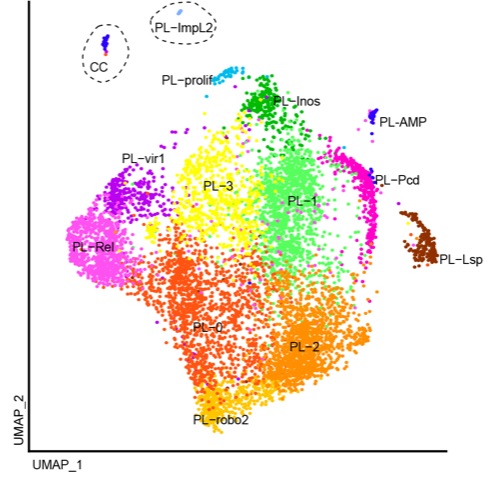
They found the presence of thirteen subpopulations of plasmatocytes and one of the crystal cells. Two populations of lamellocytes appear during the inflammatory response.
In collaboration with the team of Tina Mukherjee, Assistant Investigator at the Institute for Stem Cell Science and Regenerative Medicine (InStem, Bangalore), the analysis further showed that the hemocytes undergo a metabolic switch during development, from high production of glucose and fatty acid in the embryo to high release of energy in the larva. This change in the transcriptional landscape likely reflects the change in the environment from embryo to larvae.
Indo-French collaborative work supported by the CEFIPRA grant (Project 5903-1) sets the foundation to study macrophage heterogeneity. In the long term, these data will foster new studies on the role of the environment on macrophage biology, on the factors that drive their heterogeneity, and on the impact of specific macrophage populations in homeostasis as we all as in pathology.
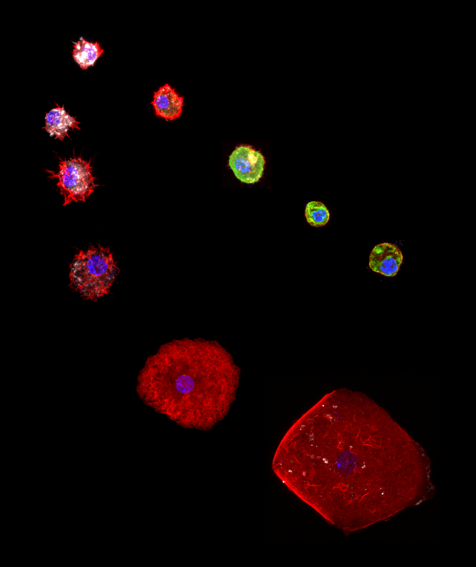
Figure 1: Different types of hemocytes found in the hemolymph of the Drosophila larva. The images were acquired with a confocal microscope and mounted using Fiji. The plasmatocytes (= macrophages) express the phagocytic receptor NimC1 (in white). They can differentiate into crystal cells expressing the marker Lozenge (in green), or upon wasp infestation, they transdifferentiate into lamellocytes strongly labelled with phalloidin (in red).
Figure 2: UMAP plot of the single-cell transcriptomic analysis of the hemocytes. The analysis reveals 13 subgroups of plasmatocytes (PL) displaying distinctive molecular signatures and one group of crystal cells (CC). The UMAP plot is a graphical representation of the single-cell transcriptomic data where each single plot represents one cell, the cells displaying similar molecular signature being grouped together. Each subgroup was label with a different color.

French PI: Dr Angela Giangrande, Directeur de Recherche CNRS, Institut de génétique et de biologie moléculaire et cellulaire, Strasbourg, France
Indian PI : Dr. Tina Mukherjee, Metabolic And Systematic Control Of Stem Cell Development, Institute for Stem Cell Science and Regenerative Medicine, Bangalore
")


")


